Test Complete
- Questions
- Score
- Minutes
| Overall Results | |
|---|---|
| Total Questions |
| Category Results | |
|---|---|
Ventilation/Perfusion (V/Q) Ratio and Mismatch
The V/Q Ratio
In respiratory physiology, the V/Q ratio refers to the ratio of ventilation to perfusion.
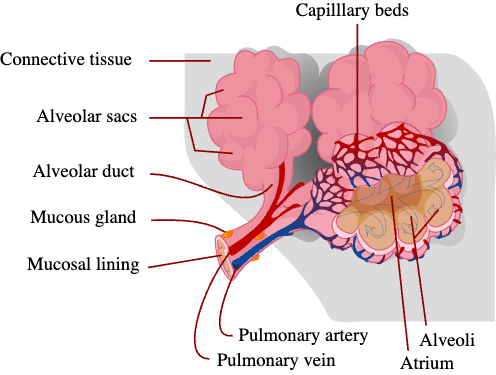
V = the amount of air that reaches the alveoli through the alveolar duct
Q = the amount of blood that reaches the alveoli through the capillary beds
In the normal lung, the V and the Q are not equal. The normal ratio is about 0.8. This is due to two main reasons: gravity and air. There is more air at the top of the lung and more blood at the bottom of the lung (because of gravity). This means that some of the blood in the bottom of the lung is not oxygenated, and some of the air in the top of the lung does not have its oxygen extracted. This concept is critical. Disruptions of V and Q are how pulmonary embolisms, pneumonia, and other lung pathologies kill patients.
Normal V/Q Values and V/Q Ratios
A normal V (alveolar ventilation)value is around 4 L/minute.
A normal Q (perfusion)value is around 5 L /minute.
Therefore, the Normal V/Q ratio is 4/5 or 0.8.
When the V/Q is > 0.8, it means ventilation exceeds perfusion. Blood clots, heart failure, emphysema, or damage to the pulmonary capillaries may cause this.
When the V/Q is < 0.8, it means perfusion exceeds ventilation. Things that may cause this are aspiration, blockage of bronchi by a foreign object, pneumonia, severe asthma, pulmonary edema, or COPD.
Apices vs Lung Bases V/Q
APICES (top): When a person is standing, the apices of the lungs have a higher V/Q ratio than the bases of the lungs. This is due to the effect of gravity on blood. The apices of the lungs are the area most superior of the organ, stretching to about the area just above the sternal end of the first rib.
BASES: The bases of the lungs are broad and concave. They sit upon the convex surface of the diaphragm, separating the left lung from the stomach, spleen, and left lobe of the liver, and the right lung from the right lobe of the liver. When standing the bases have a lower V/Q ratio than the rest of the lung, this is because the blood that is pulled down by gravity compresses the alveoli and make them harder to inflate.
Shunted Areas
SHUNTED AREA: A shunted area is an area with perfusion (Q) but no ventilation (V).
DEAD SPACE: the opposite of a shunt, dead space is an area with ventilation (V) but no perfusion (Q).
These extremes are mainly hypothetical; legitimate V/Q ratios in the areas between the shunted areas and the dead spaces are what are used clinically.