Test Complete
- Questions
- Score
- Minutes
| Overall Results | |
|---|---|
| Total Questions |
| Category Results | |
|---|---|
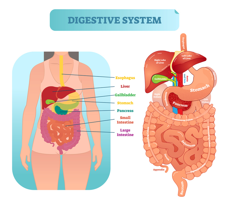
The Alimentary Tube and Accessory Organs
The Alimentary Tube and Accessory Organs
The major divisions of the digestive system include the alimentary tube and the accessory organs.
The alimentary tube functions in the digestion and absorption of food and the elimination of solid waste and begins at the mouth and ends at the anus.
Alimentary Tube Wall
There are four layers of the alimentary canal wall, including the
- inner mucosal layer, the
- submucosa, the
- external muscle layer, and the
- serosa (outermost).
Alimentary Tube Divisions and Functions
-
ESOPHAGUS: The esophagus is a muscular tube originating in the oropharynx and lying posterior to the trachea. It passes "boluses" or spherical collections of food through rhythmic muscular contractions known as peristalsis. At the base of the esophagus, there is a muscular sphincter that forms the division between it and the stomach.
The esophagus crosses the diaphragm through the ESOPHAGEAL HIATUS (at the level of T10), from the thoracic cavity to the abdominal cavity. The vagal nerves also pass through this. -
STOMACH: The stomach is located in the left upper abdominal quadrant and further macerates (and does some amount of digestion) of food that has been macerated by the teeth and tongue.
A solution of acid and enzymes in combination with physical motion provided by contractions of the stomach wall serve to break food down into a mostly liquid form known as "chyme."
-
SMALL INTESTINES: a hollow tubular organ that is located in both of the lower abdominal quadrants that is responsible for 90% of the body's food absorption capacity. It is divided into three parts, the
-
duodenum,
-
jejunum, and
-
ileum. The ileum is notable for being the site of the appendix and the ileocecal valve, a transition point to the large intestine.
DUODENUM: The duodenum begins at the exit of the stomach and is defined by its large quantity of chemoreceptors and the openings that allow liver bile and pancreatic enzymes into its lumen. The duodenum is often considered the first third of the small intestine, despite its vastly different structure. The major part of digestion takes place in the duodenum.
-
LARGE INTESTINE: Responsible for the other 10% of food absorption, the large intestine differs from the small intestine in that it mainly functions to absorb as much moisture from the stool as possible before its expulsion.
Alternately, in times of overhydration, it is one of the body's methods of eliminating water.
-
RECTUM: The last several inches of the alimentary tube is made up of the rectal vault, which stores and shapes stool for expulsion. The act of expulsion is mediated by parasympathetic neurons acting at both the rectal vault "to propel stool" and the rectal sphincters.
DEFECATION: Involves Voluntary and Involuntary Muscles.
Continence is the ability to keep feces in the rectum, made possible by the internal sphincter (under involuntary parasympathetic control) and the external sphincter, under voluntary control. The parasympathetic nerves call the shots for the internal sphincter by maintaining or changing its muscle tone, relaxing the internal sphincter when stimulated by pressure in the rectum. In fact, the internal sphincter is the major contributor (85%) to continence and the initiator of defecation with its relaxation.
Defecation: both the internal (under parasympathetic control) and the external (under voluntary control) contribute to continence and the act of defecation, but the internal sphincter relaxation under parasympathetic control initiates defecation. From that action, the external sphincter then contracts to push the feces from the anus.
Accessory Digestive Organs
Accessory digestive organs help with digestion but are not part of the digestive tract and include the
- tongue,
- salivary glands,
- pancreas,
- liver, and
- gallbladder.
TONGUE: Beyond it's role in speech, the tongue is vital for pushing food into the rear teeth and allowing them to grind it into smaller, more easily digestible pieces. It also creates the "bolus" or oval shaped collection of food that is ideal for transport via peristalsis.
SALIVARY GLANDS: These glands, located inferiorly, laterally, and superiorly in the oropharynx, serve to keep the mucous membranes of the mouth moist, prevent bacteria from destroying the teeth, and help to break down starch in the diet via the enzyme, amylase.
PANCREAS: has both an exocrine and an endocrine function. EXOCRINE: The pancreas is found in the left upper quadrant and produces enzymes that assist with digestion, specifically in the breakdown of proteins and starches. It does this by releasing these enzymes into the duodenum in response to chemoreceptors in the duodenum detecting the presence of undigested chyme. ENDOCRINE: It also keeps blood glucose levels in homeostatic ranges by controlling the release of insulin and glucagon.
LIVER: The liver is found in the upper right abdominal quadrant and processes the nutrients absorbed from the small intestine via blood flow through the portal venous system. EXOCRINE: It has the exocrine function of bile production, which serves to emulsify dietary fat and prepare it for absorption. Bile is stored in the gallbladder and is released by stimulation of the parasympathetic nervous system in response to duodenal chemoreceptors. ENDOCRINE: the liver releases factors related to glucose metabolism and growth factors.
GALLBLADDER: The gallbladder is found in the right upper abdominal quadrant and acts as a storage center for bile until needed. It is connected to the biliary tract by a small branch off of the hepatic bile duct. Bile exits the liver/gallbladder through the "common bile duct" that also carries pancreatic enzymes into the duodenum.