Test Complete
- Questions
- Score
- Minutes
| Overall Results | |
|---|---|
| Total Questions |
| Category Results | |
|---|---|
Advanced Lower Airway Structures
Category: Airway
Topic: Airway Anatomy
Level: AEMT
Next Unit: Structural Support of the Airway
7 minute read
Advanced Lower Airway Structures
The lower airway generally takes on lower importance in EMS calls than the upper airway. Obstruction and airway compromise generally occur in the upper airway, while the lower airway is usually more prone to less dramatic and more subacute processes. Knowledge of the lower airway anatomy is still important as it becomes relevant in many cardiac and systemic diseases.
The lower airway tract is defined as any airway organ below the larynx. This includes the trachea, carina, bronchi, bronchioles, alveoli, and pulmonary capillaries.
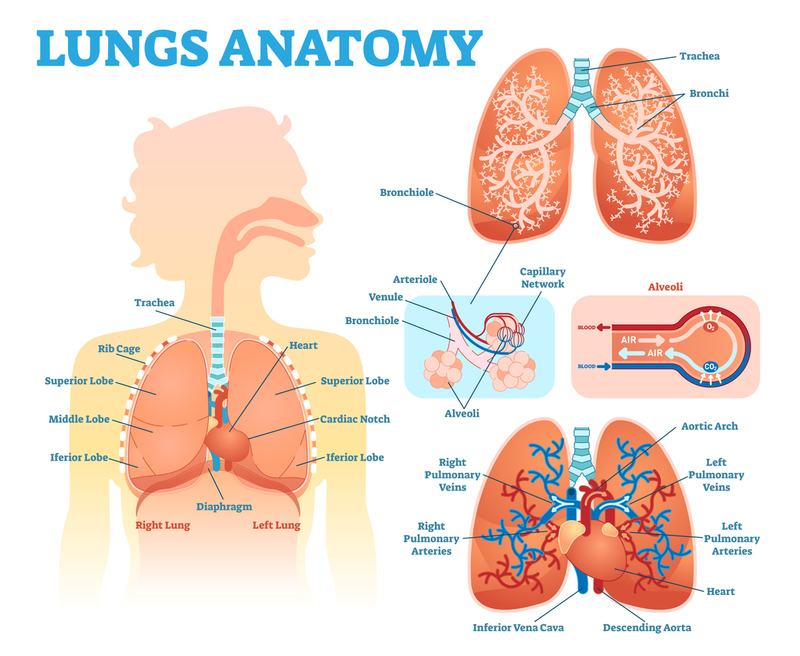
Trachea
The trachea/"windpipe" is a hollow tube that passes air to the lower airways and is supported by semicircular cartilaginous rings. The tissue of the trachea is mucosal, with hairlike cilia that push mucus, fluid, and contaminants up and out through the larynx.
Pulmonary cilia are minute hair-like organelles found in the bronchioles that move in a wavelike motion to capture, trap, and repel foreign material like dust, dirt, and smoke allowing for smooth irritant-free breathing.
The trachea passes through the mediastinum, in front of the esophagus.
Bronchi
The bronchi are hollow cartilage ringed tubes that branch off of the trachea at the carina (the bifurcation point of the trachea). They further divide to provide each lobe of the lung with a main bronchus. The right bronchus takes a straighter course at the bifurcation whereas the left bronchus is at more of an angle. For this reason, the right lung is more likely to trap foreign material that is aspirated down the trachea.
Bronchioles
The bronchioles of the lung branch off of the bronchi. They differ from the preceding elements of the airway in that they have no cartilage rings to keep them open. They instead rely on smooth muscle coated in Beta 2 receptors which allow them to change in size in response to nervous system stimulation or medication (such as albuterol and epinephrine).
Alveoli
Alveoli are the millions of thin-walled sacs surrounded by capillary blood vessels and are the site where oxygen and carbon dioxide (waste) are exchanged in the blood; pulmonary capillary beds are blood vessels that begin as capillaries surrounding each alveolus and with adequate blood volume and blood pressure, return oxygenated blood to the heart. The alveoli are considered the end of the airway.
ATELECTASIS: To stay open and continue exchanging gases the alveoli rely on the natural elasticity of the lung in combination with surfactant (a soap-like substance) secreted by the mucosal cells. The loss of this elasticity or surfactant can lead to respiratory collapse, called atelectasis, as is seen in the surfactant deficiency of immature, preterm babies. It can also occur with inadequate inflation of the alveoli, such as after abdominal surgery when deep breathing or ventilatory effort is painful.